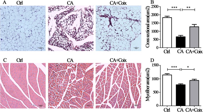
Fig. 3.
CSO attenuated muscle fiber atrophy and adipose tissue wasting induced by cancer cachexia. a. Representative figures of HE staining of epididymal adipose tissue in three groups (Ctrl, CA, CA + Coix). b.The quantitative comparison of cross-sectional area (um2) of adipocytes between three groups (Ctrl, CA, CA + Coix) revealed that CSO administration counteracted adipose tissue wasting. c. Representative cross-sectional histological staining of muscle fiber in three groups (Ctrl, CA, CA + Coix). d.The quantification of myofiber cross-sectional area showed that CSO administration attenuated muscle fiber atrophy. Values were expressed as mean ± SEM (*P<0.05, **P<0.01, ***P<0.001)

