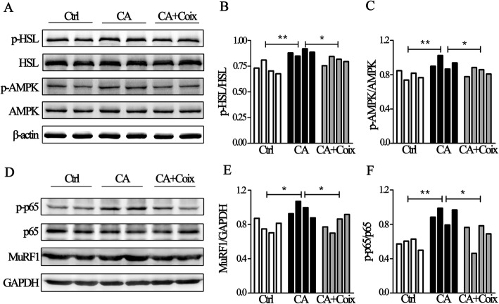
Fig. 4.
The NF-κB and AMPK pathway were involved in anti-cachectic effects of CSO. a. Representative Western-blot images exhibit the expression of p-HSL (Ser660), HSL, p-AMPK (Thr172), AMPK inthe Ctrl group, CA group, and CA + Coix group. b.The band densityanalysis showed that p-HSL/HSL in adipose tissue differed significantly between three groups (4 samples per group). c.The quantification of band density showed that p-AMPK/AMPK differed significantly between three groups (4 samples per group). d.Representative Western-blot images exhibit the expression of phospho-p65 (Ser536), p65 and MuRF1 inthe Ctrl group, CA group, and CA + Coix group. e.The quantification of band density revealed the increased ratio of MuRF1 to GAPDH in muscle tissue induced by cachexia was decreased by CSO administration (4 samples per group). f. The band densityanalysis exhibited that phospho-p65/p65 was elevated in CA group compared to Ctrl group while CSO administration significantly decreased the ratio (4 samples per group). Values were expressed as mean ± SEM (*P<0.05, **P<0.01)

