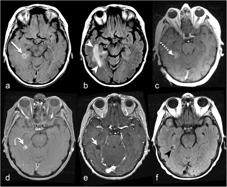
Fig. 3.

67-year-old with metastatic renal cell carcinoma. a Axial post-contrast T1-weighted imaging before LITT demonstrates a ring-enhancing metastasis in the right medial temporal lobe (long arrow). b FLAIR before LITT demonstrates surrounding vasogenic edema (white arrowheads). c Axial post-contrast T1-weighted imaging shows the ablation probe tip within the metastatic lesion (dashed arrow). d Axial post-contrast T1-weighted imaging 4 months after LITT demonstrates slightly decreased enhancement at the treated lesion (curved arrow). Axial post-contrast T1-weighted imaging (e) and FLAIR (f) 10 months after LITT show complete resolution of enhancement (short arrow) and vasogenic edema (black arrowhead)
