Fig. 4.

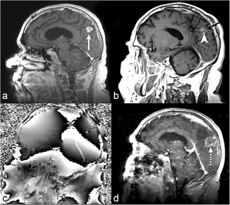
72-year-old with metastatic melanoma. a Sagittal post-contrast T1-weighted imaging before LITT demonstrates a progressing enhancing lesion in the left inferior parietal lobule at a site of a brain metastasis previously treated with gamma knife radiation therapy (long arrow). The lesion was biopsied intraoperatively immediately before LITT and was found to represent radiation necrosis. b Sagittal intraoperative localizing T1-weighted imaging with the laser probe within the lesion (arrowhead). c Sagittal intraoperative gradient-echo phase imaging is the source of the thermography maps. By subtracting subsequent images during heating from a reference image acquired before heating, a map of temperature change can be formed. d Sagittal post-contrast T1-weighted imaging 1 month after LITT demonstrates complete ablation of the lesion (dashed arrow)
