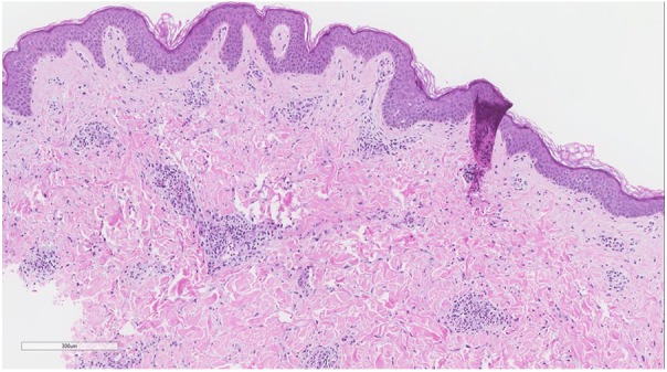
Figure 2.

Biopsy of lesional skin shows a superficial perivascular infiltrate of lymphocytes, histiocytes and occasional eosinophils. Focal, mild spongiosis is present. Subepidermal blistering is absent (Hematoxylin and eosin, 40×). Direct immunofluorescence of perilesional skin was negative (not shown).
