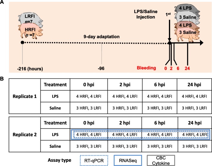
Fig. 1.
Experimental design and blood sample collection. a The experiment was performed in two replicates. Shown here is one of the two replicates. Fourteen gilts with a similar initial body weight (BW) from the low-RFI and high-RFI lines (n = 7 per line) were randomly selected and used for each replicate. Pigs were housed in individual metabolism crates, and had ad libitum access to water, but were restricted to feed intake. After a 9-day adaptation period, pigs within a line were randomly assigned to either a control (n = 6, three pigs per line) or LPS treatment (n = 8, four pigs per line) group. Pigs in the treatment group were then challenged with LPS via intramuscular injection of 30 μg/kg BW of LPS from E. coli O55:B5 dissolved in an endotoxin-free, sterile saline solution at baseline (0 hpi). Pigs in the control group were injected with an equivalent volume of endotoxin-free, sterile saline solution at the equivalent time. The rectal temperature of each pig was measured at 0, 2, 4, 6, and 24 hpi. At 0, 2, 6, and 24 hpi, blood samples were collected from the jugular vein into Tempus™ Blood RNA tubes for long-term storage at − 80 °C, into EDTA tubes for CBC tests and cytokine assays. For more details, see Materials and Methods. b Shown are the numbers of animals with blood samples collected from the two replicates at 0, 2, 6, and 24 hpi and the types of assays performed on different samples. Only blood samples collected from the LPS treated group from Replicate 2 were used for RT-qPCR and RNA-seq. Blood samples collected from all animals from both replicates were used for CBC tests and cytokine assays. The images of pigs were created by one of the co-authors, Anoosh Rakhshandeh and agreed to be published here. HRFI, high-RFI line; LRFI, low-RFI line

