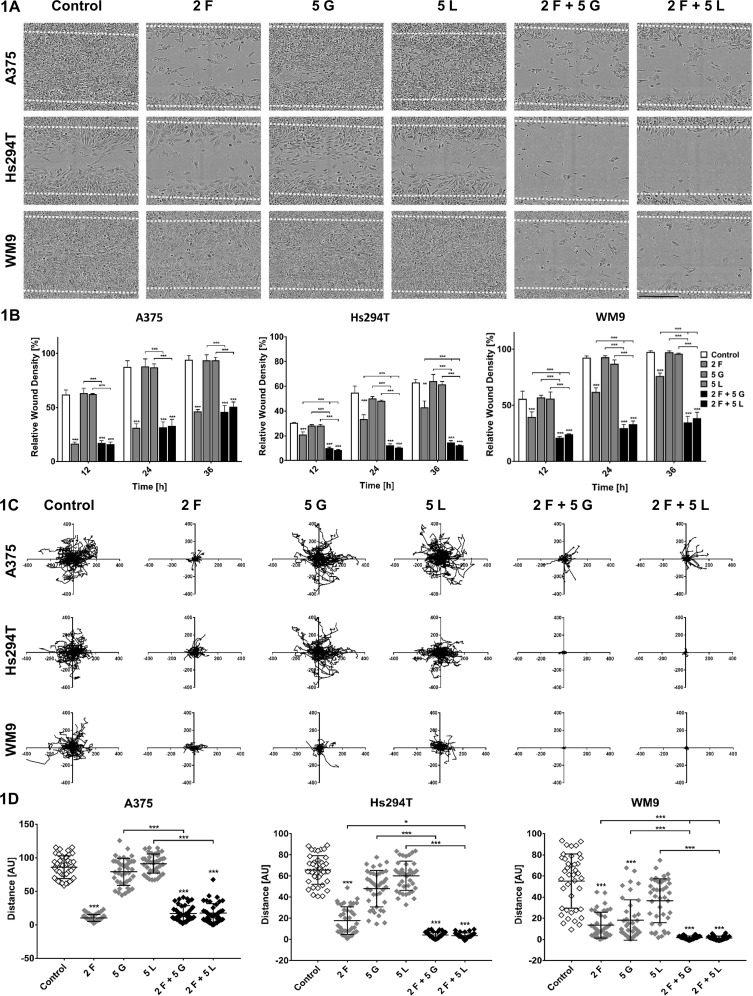
Figure 1.
Migration capacities of melanoma cells treated with inhibitors. A375, Hs294T, and WM9 cells were seeded on a thin layer of Matrigel and then incubated with foretinib [F], gefitinib [G], and lapatinib [L] or their combinations at the indicated concentrations (µM) for 36 h (A, B) or 48 h (C, D). (A) Exemplary pictures illustrating wound closure after 36 h. (B) Relative wound density was continuously measured and quantified based on pictures captured with an IncuCyte® Scratch Wound Cell Migration Software Module. (C) Cell trajectories and (D) migration distances were analyzed during 48 h of inhibitors treatment using IncuCyte® Live-Cell Analysis System and ImageJ software. Results are expressed as the mean ± SD and are based on at least three independent experiments. Asterisks indicate differences between control and treated cells or between cells treated with different drugs. The significance level was set at p ≤ 0.05 (*) and p ≤ 0.001 (***).

