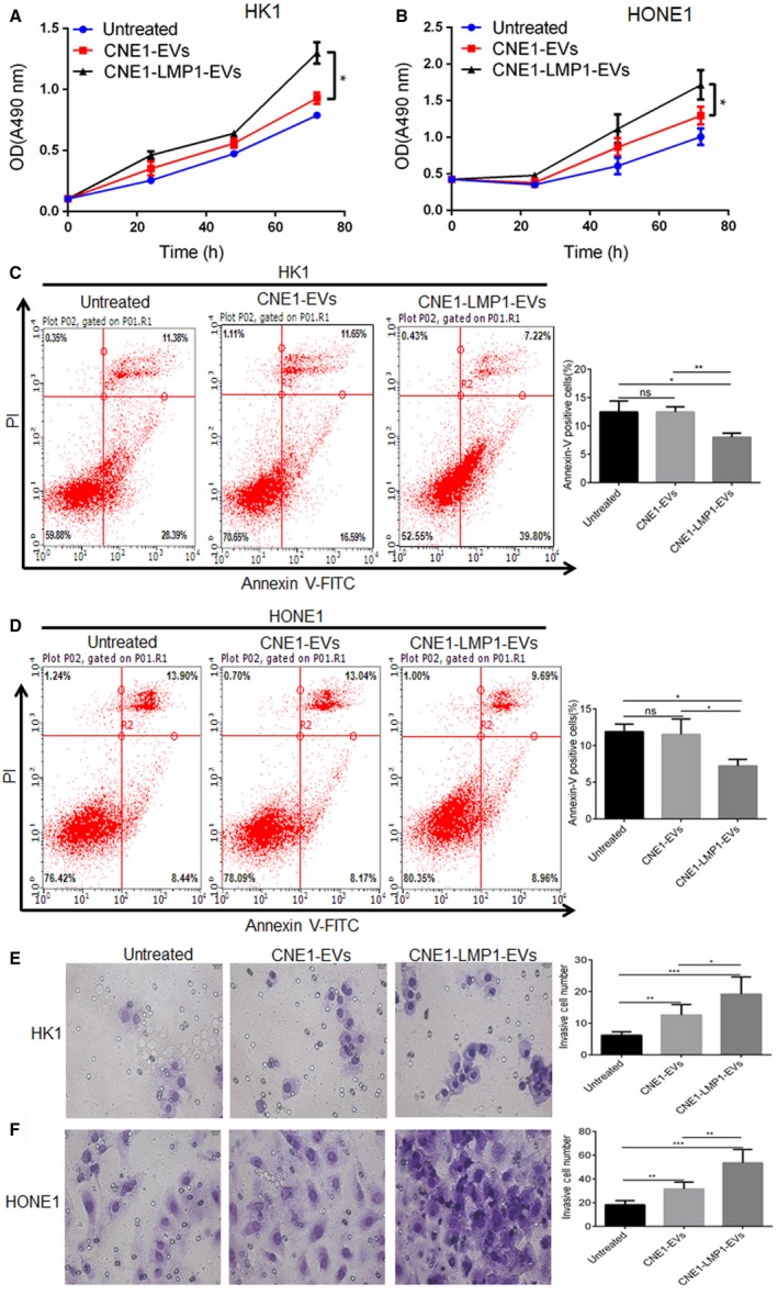
Figure 3.

CNE1‐LMP1 cell‐derived extracellular vesicles (EVs) enhance the proliferation and invasive potential and reduce the apoptosis rate of recipient nasopharyngeal carcinoma (NPC) cells. HK1 and HONE1 cells were treated with 50 μg/mL EVs from CNE1 or CNE1‐LMP1 cells for 24 h. A and B, An MTS assay was used to analyze the cell viability of HK1 or HONE1 cells after treatment with the EVs. C and D, Left: representative graphs showing the proportion of HK1 or HONE1 cells that were positively stained with Annexin‐V. Right: analysis of three independent experiments assessing the proportion of Annexin‐V‐positive cells. E and F, Left: representative graphs showing the invasion of HK1 or HONE1 cells through the 0.4‐μm pore of the transwell. Right: statistical analysis of three independent experiments assessing the number of HK1 or HONE1 cells in five visual fields. The results are presented as the mean ± SD.*P < .05, **P < .01, ***P < .001, ns not significant
