Fig. 1.
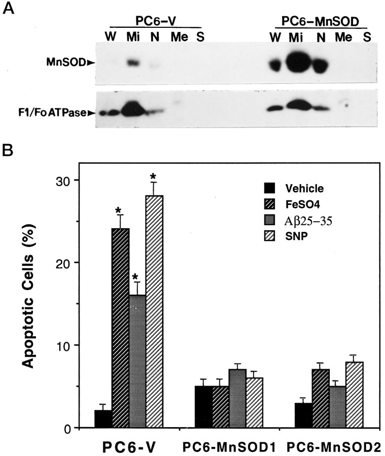
Expression of human MnSOD in PC6 cells: localization to the mitochondria and protection against oxidative stress-induced apoptosis. A, Western blot analysis and subcellular fractionation of MnSOD and F1/Fo-ATPase levels in PC6-V and PC6-MnSOD cells are shown. W, Whole cells;Mi, mitochondrial fraction; N, nuclear fraction; Me, membrane fraction; S, soluble fraction (cytosol). Fifty nanograms of protein/lane (for both PC6-V and PC6-MnSOD cells) were separated by SDS-PAGE, transferred to a nitrocellulose sheet, and immunoreacted with antibodies to either MnSOD (upper) or the mitochondrial enzyme F1/Fo-ATPase (lower). The upper and lower blots are two separate blots using the same sample preparations; the upper blot was reacted with the MnSOD antibody, and the lower blot was reacted with the F1/Fo-ATPase antibody. Note that MnSOD is localized almost exclusively in the mitochondrial fraction. B, Cultures of PC6-V and two different lines of PC6-MnSOD cells were exposed for 24 hr to saline (vehicle), 100 μm FeSO4, 50 μm Aβ, or 100 μm SNP, and the percentages of cells exhibiting apoptotic nuclei were determined. Values are the mean ± SEM of determinations made in eight cultures; *p < 0.01 compared with the value for vehicle-treated cultures and with each value in PC6-MnSOD cells (ANOVA with Scheffe’s post hoc tests).

