
Fig. 7.
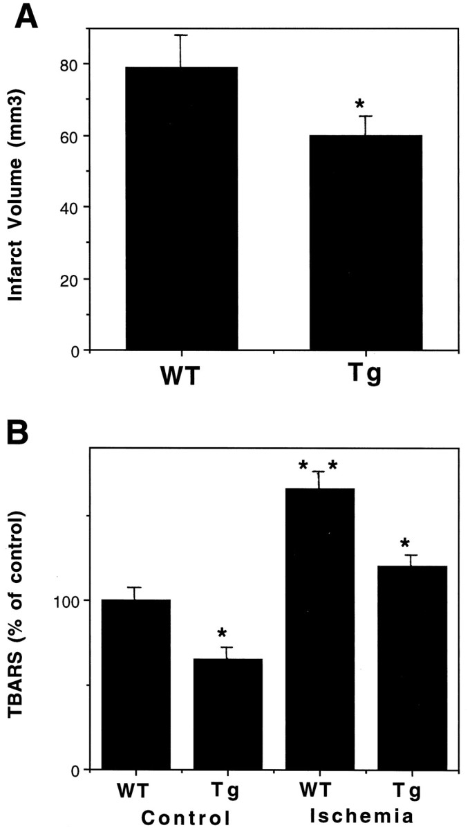
Levels of cellular injury and lipid peroxidation are reduced in MnSOD Tg mice after cerebral ischemia. A, Cortical infarct volumes were quantified 24 hr after MCA occlusion in WT and MnSOD Tg mice. Values are the mean ± SEM (n = 6 mice in each group); *p< 0.05 compared with the WT value. B, TBARS levels were quantified in infarcted cortical tissue 24 hr after MCA occlusion in WT and MnSOD Tg mice. Values are the mean ± SEM (n = 4 mice in each group); *p< 0.01 compared with the corresponding WT value; **p < 0.01 compared with the WT control value.