
Fig. 3.
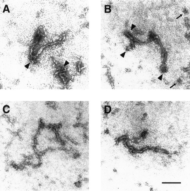
Localization of NR1 along assembled neurofilaments by immunogold electron microscopy. Purified NF-L was assembled in vitro in the presence (A,B, D) or absence (C) of NR1a C-terminal fusion protein (see Materials and Methods). Assembled filaments were adsorbed onto carbon-coated copper grids and stained with either anti-NR1 antibody (A–C) or control antisera (D) followed by gold-conjugated secondary antibody (6 nm gold particle). Labeled filaments were visualized by electron microscopy. Gold particles were observed at the ends of filaments (A, B,arrowheads) and sometimes over amorphous regions of electron density assumed to represent truncated filaments (B, thin arrows). Magnification, 224,000×. Scale bar, 0.1 μm.