
Fig. 2.
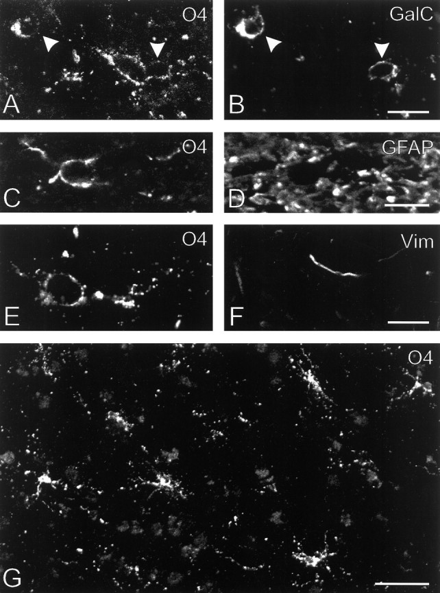
Antigenic and morphological characteristics of oligodendrocyte precursor cells in chronic MS lesions. A, B, An O4-positive, GalC-negative oligodendrocyte precursor cell and two GalC-positive, weakly O4-positive, rounded oligodendrocytes (arrowheads) that were present in the center of lesion H (subject 96-040). C, D, Although the O4-positive (GalC-negative) cells were surrounded by large numbers of GFAP-positive filaments, confocal laser-scanning microscopic analysis suggested that they themselves lacked GFAP and thus were oligodendrocyte precursor cells. Detail of lesion Q, subject 96-121. E, F, The O4-positive (GalC-negative) oligodendrocyte precursor cells also lacked the intermediate filament vimentin (Vim).G, Low-power view of an area of lesion F (subject 96-039) that contained large numbers of O4-positive (GalC-negative) oligodendrocyte precursor cells (Table 2). This lesion also contained numerous debris-laden macrophages (Table 2), and because the debris within these cells was slightly autofluorescent, they are vaguely visible in the background. Scale bars: B, D, F, 10 μm;G, 50 μm.