
Fig. 1.
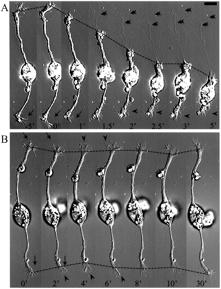
BDNF-induced collapse of growth cones of cultured embryonic Xenopus spinal neurons. Time-lapse DIC sequences showing growth cone collapse followed by dramatic (A) or moderate (B) neurite retraction induced by BDNF. Numbers represent minutes at various times before (negative numbers) and after (positive numbers) the application of 50 ng/ml BDNF (at time 0). In B, BDNF was washed out at 10 min after the application, and neurite extension was observed thereafter. Note the transient loss of filopodia (arrows) after BDNF application and the appearance of lamellipodial protrusion at later times (arrowheads). In A, also note that some adhesion remained during growth cone collapse (double arrowheads). Dashed lines depict the positions of the growth cones at various times. Scale bar, 15 μm.