Fig. 3.
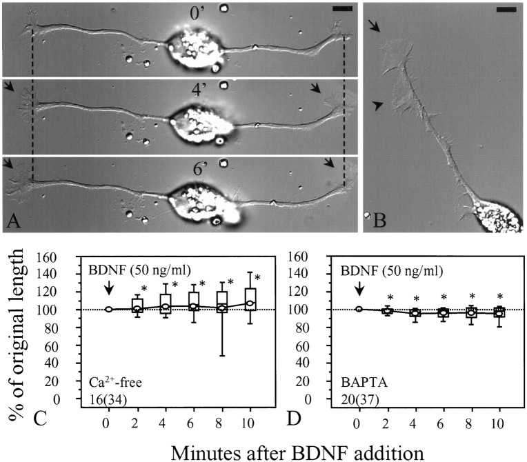
Ca2+ mediates the collapsing effect of BDNF. A, Representative images showing the changes induced by BDNF (50 ng/ml) in Ca2+-free medium. No growth cone collapse and neurite retraction were observed after BDNF addition. However, BDNF-induced lamellipodial protrusion was still observed (arrows). Numbersrepresent minutes after BDNF addition. Dashed linesindicate corresponding positions along the neurite. B, BDNF-induced lamellipodial protrusion was observed not only at the growth cone (arrow) but also along the neurite shaft (arrowhead). The image was taken 10 min after BDNF application. C, Data from populations of cells in the Ca2+-free medium are presented as a box whisker plot. Apparently no collapse was observed after BDNF application. D, Loading cells with 10 μmBAPTA AM for 20 min also blocked the collapsing effect of BDNF. No substantial changes in neurite length were observed. *Significantly different from the POL of BDNF in culture medium (Fig.2B) at corresponding times after BDNF addition (p < 0.001, Mann–Whitney test).Numbers in C and Dindicate the numbers of cells (growth cones) examined. Scale bars, 10 μm.

