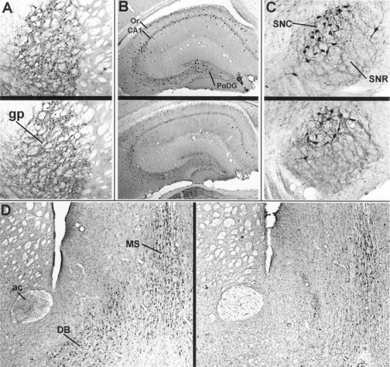
Fig. 2.

Comparison of anti-nAChRα4 immunoreactivity in five brain regions of young or aged CBA/J mice. Anti-nAChRα4 antisera reveal putative neuronal cell staining in representative brain sections taken from a young (A–C, upper;D, left) or aged (A–C,lower; D, right) CBA/J male mouse. The regions emphasized are the globus pallidus (gp) in A; the hippocampus [CA1, the polymorphic layer of the dentate gyrus, and the stratum oriens (Or)] in B; the substantia nigra [compacta (SNC) and reticulata (SNR)] in C; and the medial septum (MS), diagonal band (DB), and anterior commissure (ac) in D.
