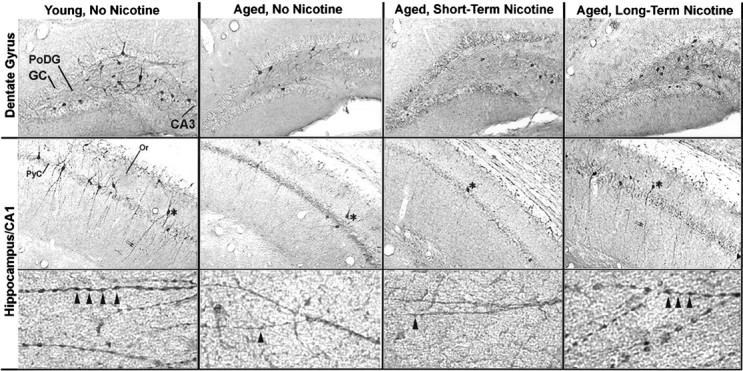
Fig. 4.

Anti-α4 immunoreactivity in hippocampal sections from representative young and aged male CBA/J mice that were grouped according to receiving no nicotine (control) or nicotine in their water for 6 weeks (short-term) or 11 months (long-term). As described in Materials and Methods, three male CBA/J mice were given nicotine and saccharin in their water for 6 weeks (24 months old at start of treatment) or 11 months (14 months old at start of treatment). Controls received water with saccharin but without nicotine. In the top row is a representative section from the dentate gyrus of each group exhibiting the immunoreactivity of neurons to anti-nAChRα4 antibodies. The arrow (left) points to a stained neuron in the polymorphic layer. Other labels show the granule cells (GC) of the dentate gyrus and neurons of the CA3. In the middle row is shown the hippocampal CA1 region for each group. The asterisks note an anti-nAChRα4-immunopositive cell in each treatment group. The pyramidal cell layer (PyC) and the Or are noted. The bottom row shows at high magnification the immunopositive processes that extend ventrally from stained cells of the CA1 (middle row). These cells exhibit extensive dendritic-like processes that exhibit regular varicosities on greater magnification (arrowheads). A typical young animal (3 months) is shown on the left; a typical older animal (25 months) is shown on the right. These cells are less abundant, and the large processes exhibiting varicosities are diminished or entirely absent in the older animals except in the aged group that was supplied with nicotine in their water for 11 months.
