
Fig. 2.
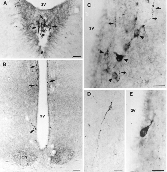
Immunohistochemical visualization of sst1 receptor in coronal sections of the rat hypothalamus.A, Organum vasculosum laminae terminalis showing sst1 labeling of nerve fibers in the ependyma (arrows). B, A strong staining for sst1 in perikarya (arrows) in the periventricular area surrounding the anterior part of the third ventricle (3V). In the suprachiasmatic nucleus (SCN), a moderate labeling of nerve fibers is observed. C, Immunostained nerve fibers (arrows) and neuronal perikarya (arrow heads) showing varying labeling intensity in the periventricular nucleus. D, In the hypothalamus, single-labeled perikarya and nerve fibers were observed.E, Micrograph showing a strongly labeled cell body positioned within the ependymal layer of the third ventricle. Scale bars: A, B, 200 μm; C–E, 50 μm.