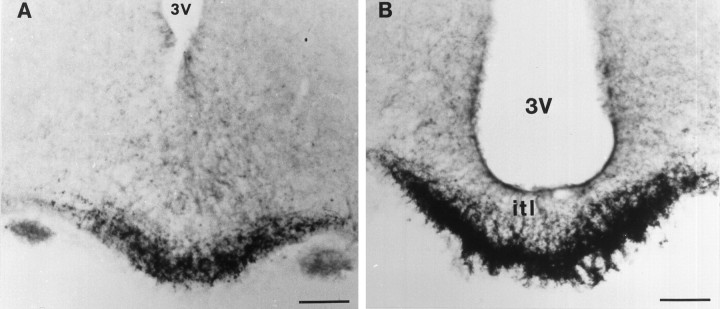
Fig. 3.
Detection of sst1 receptor immunoreactivity in coronal sections of the median eminence. An intense staining of nerve fibers endowed with large swellings is seen in the rostral part of the organ (A). In the more caudal part of the median eminence (B), the labeling is clearly confined to the external layer surrounding the portal capillaries. itl, Internal layer. Scale bars, 200 μm.

