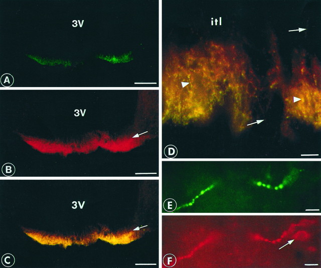
Fig. 4.
Double-immunofluorescent labeling of the rat median eminence. sst1 is visualized by fluorescein (green) and somatostatin with Texas Red (red). A strong labeling was observed in the external layer for sst1 (A) and somatostatin (B). A double-exposure micrograph is shown (C) displaying colocalization of somatostatin and sst1. Arrows mark the border between the internal and external layers of the median eminence. Double-exposure micrograph of high magnification of the median eminence (D) shows nerve fibers with boutons containing both sst1 and somatostatin (arrowheads) or somatostatin alone (arrows). itl marks the position of the internal layer. A periventricular nerve fiber endowed with large boutons shows colocalization of sst1(E) and somatostatin (F). Note the perikaryon exhibiting a positive immunoreaction for somatostatin (arrow) but not for sst1. Scale bars: A–C, 200 μm;D–F, 25 μm.

