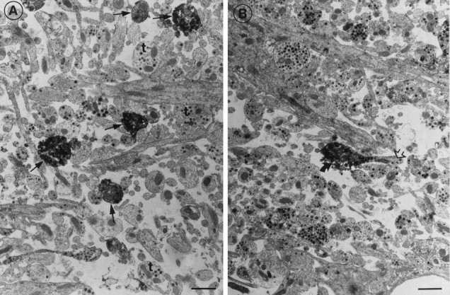
Fig. 5.

Electron microscopical detection of sst1 receptor immunoreactivity in the median eminence.A, Several immunoreactive nerve terminals (arrows) are seen between unstained nerve terminals (t). B, An unstained nerve fiber (open arrow) terminates in an immunoreactive nerve terminal (bent arrow). Scale bars, 1 μm.
