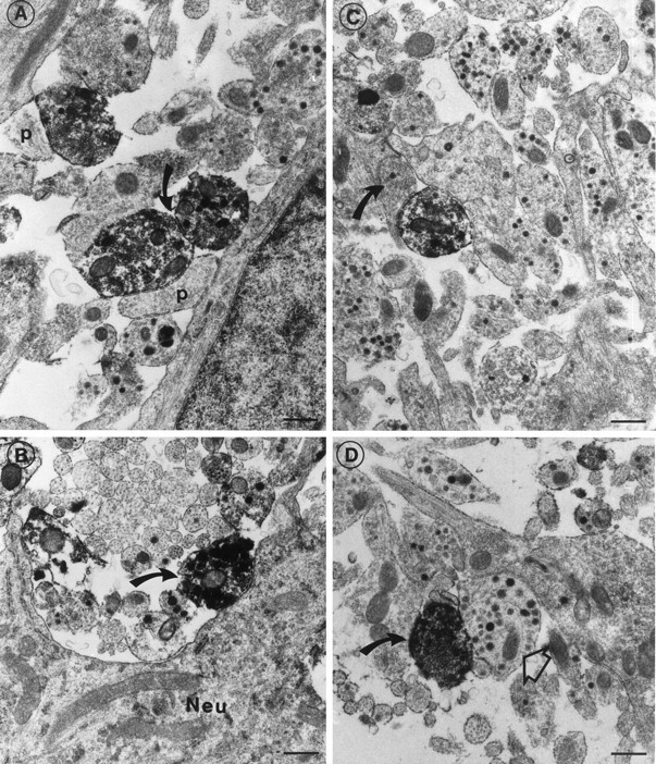
Fig. 6.

Electron micrographs of the median eminence immunoreacted for the sst1 receptor. A, Immunoreactive nerve terminals making synaptic contacts with unstained postsynaptic structures (p). Both presynaptic and postsynaptic structures contain clear neurotransmitter vesicles. Note the synaptic junction (bent arrow) between two immunoreactive terminals. B, Positive nerve terminal (bent arrow) making synapse-like contact with a neuronal perikaryon (Neu). C, Electron micrograph of an immunoreactive terminal making a synaptic contact with an unstained bouton termineaux (bent arrow) with small clear and a single large dense-core vesicle.D, Immunoreactive nerve terminal (bent arrow) with small clear and large dense-core vesicles. A classic peptidergic nerve terminal (open arrow) with many dense-core vesicles is seen close to the immunoreactive terminal. Scale bars, 0.5 μm.
