
Fig. 1.
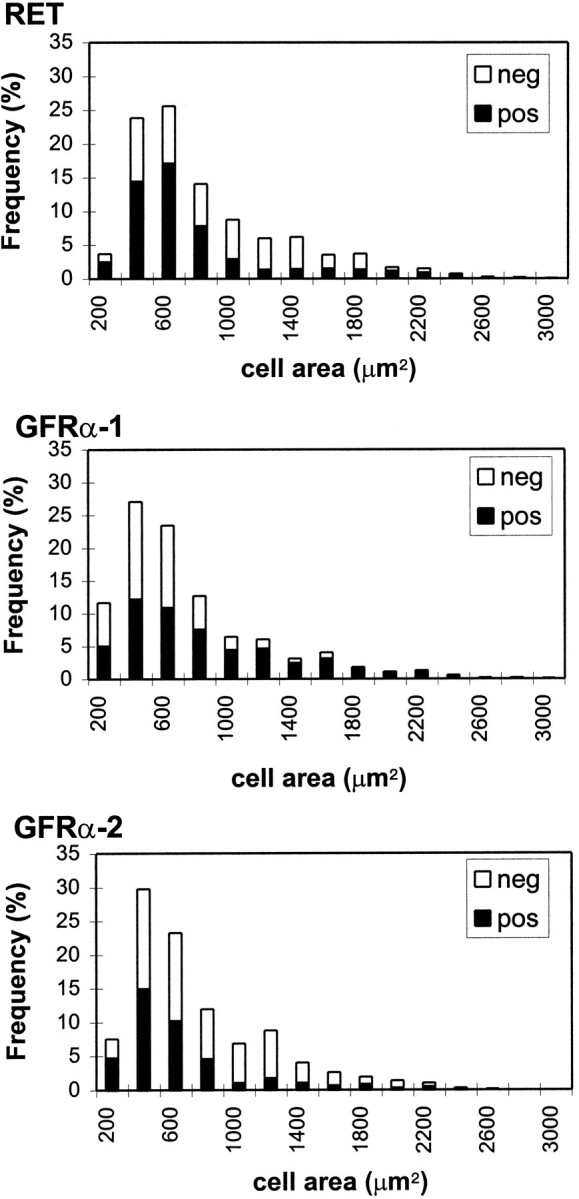
Cell size distribution of DRG cell profiles positively and negatively labeled for RET, GFRα-1, and GFRα-2 within L4/5 dorsal root ganglia. RET and GFRα-2 are present predominantly in small and intermediate diameter DRG cell profiles but are also present in some large diameter DRG cell profiles. GFRα-1 is more evenly distributed through the whole cell size spectrum.