
Fig. 4.
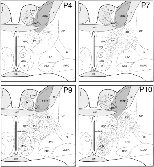
Preoptic projections of the BSTp.MPN, Illustrations show the distribution of DiI-labeled fibers at the level of the MPN in male rats perfused onP4, P7, P9, andP10. The dark gray area in the BST represents the area of DiI diffusion immediately adjacent to the implant site. BAC, Bed nucleus of the anterior commissure; DBB, nucleus of the diagonal band;GP, globus pallidus; MPNc, medial preoptic nucleus, central part; MPNl, medial preoptic nucleus, lateral part; MPNm, medial preoptic nucleus, medial part; och, optic chiasm; PD, posterodorsal preoptic nucleus; PvPo, preoptic periventricular nucleus. See Figures 2 and 3 for additional abbreviations.