
Fig. 6.
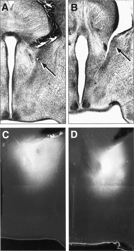
A, B, Low magnification images of Nissl-stained sections showing the precise placement of the DiI crystal in the BSTp of a P10 male (A) and a P10 female (B) rat. The arrows indicate the location of the center of the DiI implant. C, D, Low magnification, combined dark-field and fluorescence image montages showing the appearance and distribution of DiI in comparable implants obtained in P10 male (C) and female (D) rats.