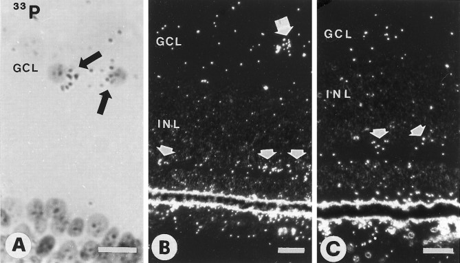
Fig. 3.
Retinae of E15 chick embryos hybridized with33P-labeled riboprobes for chicken BDNF. A, Bright-field view of two labeled cells in the ganglion cell layer (GCL, arrows). B, Dark-field image of a labeled cell in the GCL (larger arrow) and several labeled cells in the outer half of the inner nuclear layer (INL, smaller arrows).C, Dark-field image of E15 retina after optic stalk transection at E4. Label in the outer half of the INL is maintained (arrows). Scale bars: 10 μm in A; 20 μm in B, C.

