
Fig. 6.
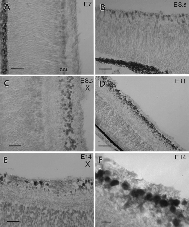
BDNF immunoreactivity in the retina.A, The ganglion cell layer (GCL) of an E7 retina is weakly labeled. B, At E8.5 newly formed cells adjacent to the ora serrata are BDNF-positive. These cells presumably represent retinal ganglion cells (RGC). C, In an E.8.5 retina BDNF is not visibly reduced in the GCL after optic stalk transection (X) at E4 (compare with Fig.5A). D, At E11 many cells in the normal GCL are strongly labeled. E, After optic stalk transection (X) many RGC have degenerated at E14. Some large cells display strong BDNF immunoreactivity.F, An E14 retina labeled with the BDNF antiserum at higher magnification displays heterogeneity of the label within the GCL. Scale bars: 25 μm in A–C, E; 50 μm in D; 10 μm in F.