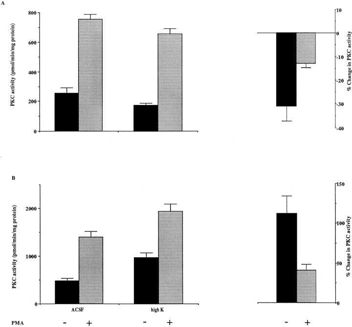
Fig. 6.
The effects of high-K ACSF treatment on PKC activity of P8 (A) and adult (B) cerebellum. P8 and adult cerebellar slices were incubated in ACSF or high-K ACSF for 2 hr. PKC activity of homogenates was determined in the presence or absence of 10 μm PMA. Changes in PKC activity were calculated according to the following equation: (PKC activity (high K)/PKC activity (ACSF) −1) × 100%. The PMA-independent PKC activity in control (ACSF) sample at P8 is significantly different from that in the high-K treated sample; p < 0.05, by a one-tailed Student’st test. The interaction between age of the animal and the effects of high K treatment is significant; p< 0.05 for PMA-independent PKC activity; p < 0.01 for the PKC activity in the presence of PMA, using a two-factor ANOVA test (n = 4). The PKC activity in adult high-K-treated sample is significantly different from that in the adult control (ACSF) and in the P8 high-K-treated sample;p < 0.01 (without PMA) and p< 0.01 (with PMA). PKC activity in the adult control is different from that in P8 control; p < 0.05 (without PMA) andp < 0.01 (with PMA), by a Tukey’s multiple-comparisons test. The depolarization-induced change in PKC activity in the absence and presence of PMA in the adult is significantly different from that at P8; p < 0.001 (without PMA) and p < 0.01 (with PMA), by a two-tailed Student’s t test.

