
Fig. 5.
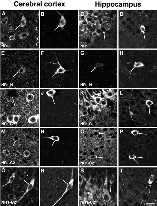
Localization of NMDAR1 isoforms in the cerebral cortex and hippocampus. The left two columns illustrate neurons in the cerebral cortex, within lamina III–V. The right two columns illustrate the CA1 region of the hippocampus. In each of the illustrations of the hippocampus, the stratum pyramidale is in the upper left and the stratum radiatum is in thelower right part of each panel, except for panelsS and T, where the orientation is the same but only the stratum pyramidale can be seen. In each case, staining for NMDAR1 is illustrated on the left, whereas staining of the same region for nNOS is on the right. The antibody to the conserved region of NMDAR1 produces strong staining of numerous cortical (A) and hippocampal (C) neurons; in both regions the nNOS neurons are labeled (B, D). The antibody to the N1 segment produces some low-intensity labeling of cortical nNOS neurons (E, F) but only very sparse labeling in the hippocampus (G,H). The antibody to the C1 segment stains cortical (I) and hippocampal pyramidal (K) neurons intensely, but it does not stain the nNOS cells in these regions (I,J and K, L). As in the striatum, the C2 segment antibody stains the cytoplasm of neurons as well as the neuropil in the cortex (M, N) and hippocampus (O, P); this staining is similar in nNOS-positive and nNOS-negative neurons. The antibody to the C2′ segment stains a moderate number of cortical neurons (Q) and all of the pyramidal cells of CA1 (S). In both regions the nNOS neurons are strongly stained (Q, R and S, T) for the C2′ segment. Scale bar, 20 μm.