
Fig. 7.
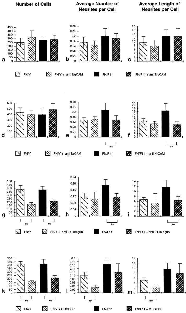
Perturbation of F11-induced enhancement of neurite outgrowth by different reagents. Retinal cells were cultured on an FN–IgY or an FN–F11 substrate in the presence or absence of polyclonal NgCAM-specific Fab fragments (a–c), NrCAM-specific Fab fragments (d–f), a β1-integrin-specific mAb (g–i), or GRGDSP peptides (k–m). Cultures were evaluated, and data are presented as outlined in the legend of Figure 2. Data from one representative experiment of at least three independent experiments are shown. **Statistical significance of the difference (Mann–WhitneyU test, p < 0.001). The number of substrate-bound cells was not influenced by application of the NrCAM or NgCAM antibodies (a, d). On the FN–IgY substratum, neither the NgCAM antibodies (b, c) nor the NrCAM antibodies (e, f) interfered with the average number of neurites per cell (b, e) or the average length of neurites per cell (c, f). On the FN–F11 substratum, the NgCAM antibodies also did not interfere with the F11-induced enhancement of outgrowth (b, c). By contrast, the NrCAM-specific antibodies reduced outgrowth to the FN-dependent level (e, f). The β1-integrin antibody (g) and the GRGDSP peptide (k) interfere significantly with cell attachment on both substrates. Although the antibody did not reduce FN-dependent basal neurite outgrowth (h, i), the peptide strongly decreased outgrowth on the FN substrate (l, m). In the presence of the antibody, the subpopulation of attached cells does not respond to F11 in the FN–F11 substrate but shows only FN-dependent basal outgrowth (h,i). By contrast, the cells that are attached in the presence of the peptide show F11-mediated enhancement of outgrowth, which is not significantly different (p > 0.001) from that in the absence of the peptide (l, m).