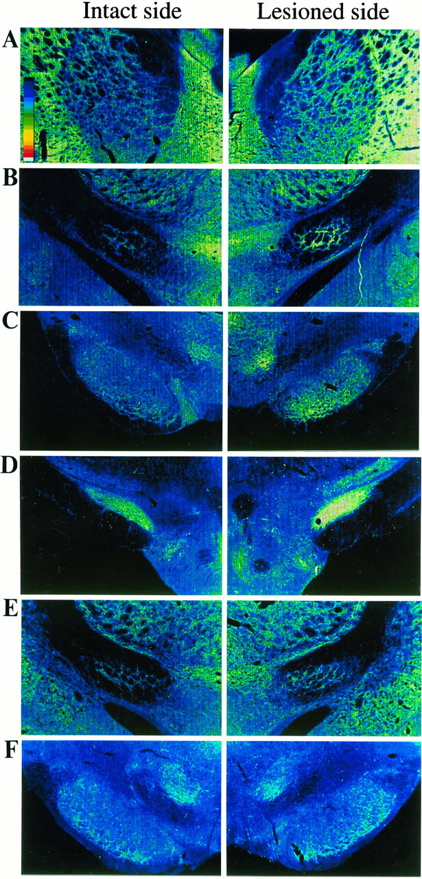
Fig. 4.

Digitized images of cytochrome oxidase (CO) activity in basal ganglia nuclei. In rats with unilateral nigrostriatal lesions, increased optical densities are noted in the GP (A), EP (B), SNR (C), and STN (D) ipsilateral to the lesion. In the lesioned animals with nigral grafts, the increases in CO activity are attenuated in the EP (E) and SNR (F).
