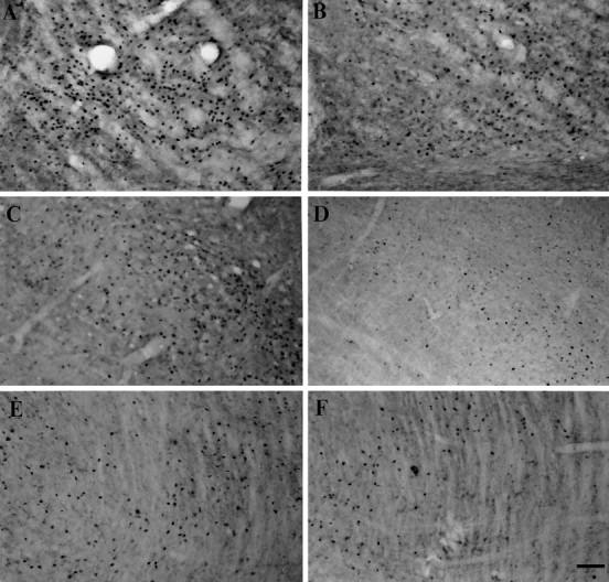
Fig. 7.

Photomicrographs showing c-Fos-immunopositive cells in the VM (A, B), SC (C, D), and PPN (E, F) on the lesioned side after systemic administrations of apomorphine. In the lesioned animals, the numbers of c-Fos-positive cells are increased in the VM (A), SC (C), and PPN (E) on the lesioned side after systemic apomorphine challenge. The apomorphine-induced enhancement of c-Fos expression in these structures is attenuated in the lesioned rats with nigral grafts (B, D, F). Scale bar, 100 μm.
