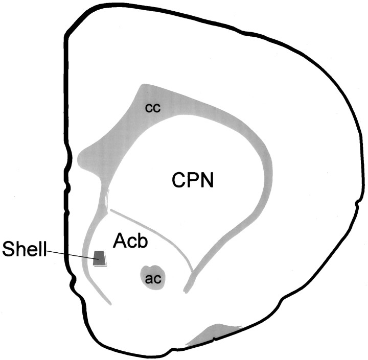
Fig. 1.
Coronal hemisection of the rat forebrain. Schematic diagram illustrating the region of the Acb shell that was sampled for electron microscopic analysis. ac, Anterior commissure; Acb, nucleus accumbens; cc, corpus callosum; CPN, caudate–putamen nucleus. Modified from Swanson (1992).

