
Fig. 4.
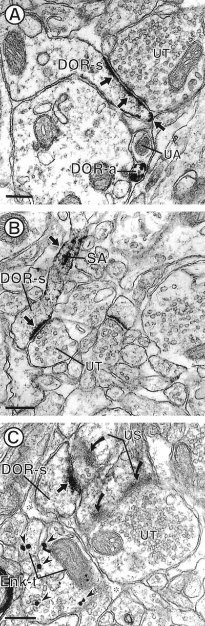
Electron micrographs showing DOR immunoreactivity in dendritic spines and their relationship with Enk-labeled terminals. The dendritic spines in A andB (DOR-s) show intense DOR-LI mainly localized to plasma membranes of postsynaptic densities and along nonsynaptic portions of dendritic spines (small arrows).B, DOR-LI within the spiny apparatus (SA) and along the plasma membrane of a spine neck (small arrow). Both DOR-labeled spines receive asymmetric synaptic input from unlabeled terminals (UT).C, Dendritic spine (DOR-s) that has intense DOR labeling along the postsynaptic density (small arrow). DOR-s receives synaptic input from an unlabeled terminal that also contacts an unlabeled spine (US). DOR-s is also apposed to two axon terminals (Enk-t) that contain gold–silver particles for Enk (arrowheads). Enk-t are separated from DOR-s by glial processes (asterisks). The unlabeled terminal also forms a synapse with another spine (US), which lacks DOR immunoreactivity. In the same field, an unlabeled spine (US) forms a perforated asymmetric synapse (curved arrows) with an unlabeled terminal (UT). Scale bars, 0.26 μm.