
Fig. 5.
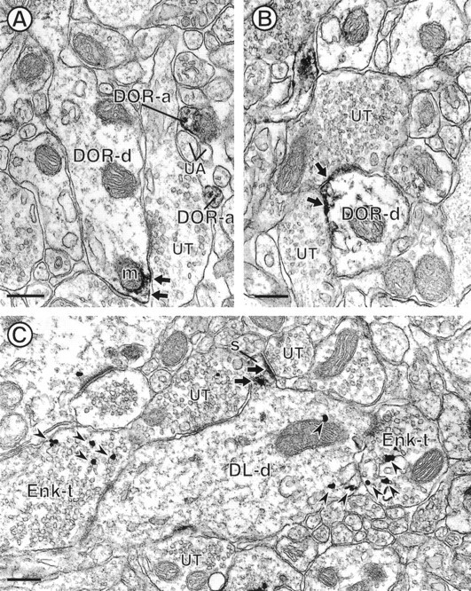
Electron micrographs showing DOR-LI in dendrites and their relationship to Enk. The dendrites in A andB (DOR-d) show peroxidase reaction product for DOR (small arrows) along selective portions of their plasma membranes. Diffuse reaction product is also seen along the membranes of nearby cytoplasmic organelles, including a mitochondrion (m) in A. Both of the DOR-labeled dendrites are apposed to unlabeled terminals (UT). Within A, there are also two small unmyelinated axons that are labeled for DOR (DOR-a) within a group of unlabeled axons (UA). C, Dendrite that is dually labeled for DOR and Enk (DL-d). The dendritic spine (S) is intensely labeling for DOR (small arrows), whereas the gold–silver particles for Enk are contained within the shaft (arrowheads).DL-d receives convergent input from two Enk-immunoreactive terminals (Enk-t) and two unlabeled terminals (UT). Scale bars, 0.27 μm.