Fig. 1.
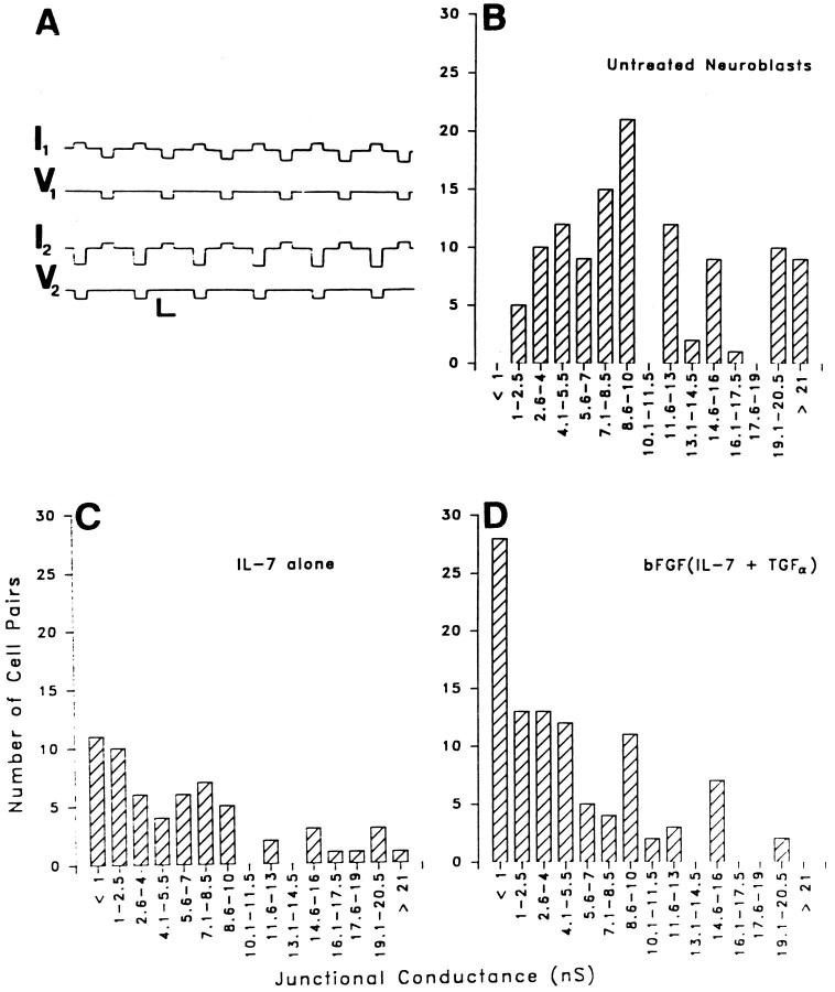
Junctional conductance measurements revealed that coupling strength is higher in the untreated neuroblasts than in cells that differentiated in response to either treatments. Untreated and cytokine-treated neuroblasts were cultured for the same length of time (1–7 DIV). A, Recording of macroscopic junctional conductance in a pair of untreated neuroblasts. Command voltage pulses were applied alternately to cells 1 and 2. Currents recorded in the same cell in which the voltage step is applied represent the sum of conductances of junctional (gj) and nonjunctional (gnj) membranes; junctional currents are recorded in the other cell. Calibration bars: horizontal, 1 sec; vertical, 10 mV (V1,V2), 50 pA (I1,I2). B, Histogram of junctional conductance (gj) values obtained in 115 pairs of untreated neuroblasts. The mean value of gj was 11.28 ± 0.7 nS (median, 10 nS). C, Histogram of gjvalues obtained from neuroblasts treated with IL-7 alone (n = 60). Mean gj was 6.51 ± 0.77 nS (median, 5 nS). D, Histogram ofgj obtained in 100 pairs of neuroblasts treated with bFGF(IL-7 and TGFα). Mean gjwas 5.07 ± 0.51 nS (median, 4 nS).

