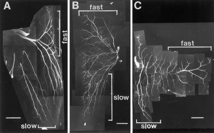
Fig. 1.
Intramuscular nerve branching patterns of three hindlimb muscles with separate fast and slow muscle regions. Muscle whole mounts of IFIB (A), SART (B), and AITIB (C) at stage 36 were stained with anti-neurofilament antibody to visualize the nerves. A–C, Anterior is to theleft, proximal is at the top, and myotubes are oriented top to bottom.A, Nerves in the slow region of the IFIB grew parallel to the myotubes (lower bracket) and sent off multiple collateral sprouts at regular intervals. In contrast, axons in the fast region grew transversely across the myotubes before branching in a “reductive” pattern to innervate focally the myotubes (upper bracket). B, Motor axons entered the SART at the posterior muscle edge and grew proximally to reach the fast region (upper bracket) or distally to innervate the slow region (lower bracket). Despite the proximal and distal orientation of the muscle regions, axon branching patterns resembled those in the IFIB. Axons innervating the slow SART grew predominantly along the myotubes (lower bracket), sending off small branches, whereas axons in the fast region first grew across the myotubes and branched reductively (upper bracket).C, Axons in the slow AITIB (lower bracket) entered the muscle and grew longitudinally along the muscle fibers, sending off multiple branches similar to those in the IFIB. In the fast region (upper bracket), axons grew perpendicular to the myotubes and then branched sparsely in a reductive pattern. Scale bars: A, B, 500 μm;C, 1 mm.

