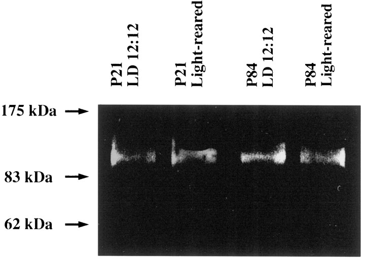
Fig. 7.
GluR1 in rat retina demonstrated by immunoblotting. Total tissue homogenates (P21 or P84) from rat retina (20 mg/lane) were electrophoresed by SDS-PAGE and transferred to nitrocellulose membrane. Immunoblotting was performed using antibodies specific for GluR1. A single immunoreactive band appeared at ∼100 kDa. The density of the band was almost identical in two different lighting conditions at both P21 and P84 as quantified by CCD imaging.

