
Fig. 6.
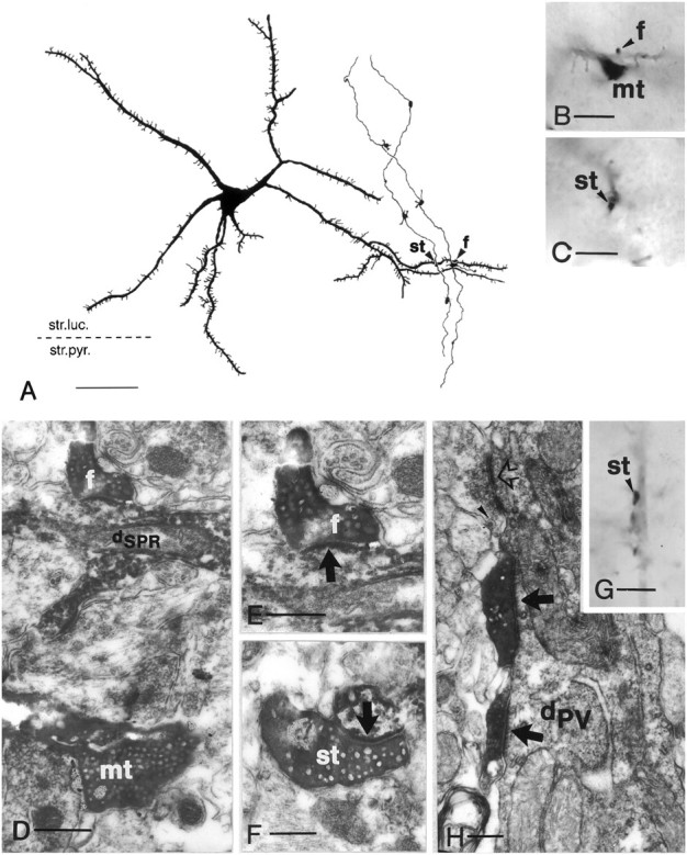
Convergence of adjacent granule cells to a spiny SPR-immunoreactive cell (A–F) and innervation of a parvalbumin-positive interneuron by a granule cell (G–H). A, Camera lucida drawing of an SPR-positive spiny neuron in the CA3c subregion (neuron3 in Fig. 4B). The same tertiary dendrite is contacted by mossy fibers of two neighboring granule cells (arrowheads). The left contact is formed by a smallen passant terminal (st), whereas the right contact is formed by a filopodial extension (f). B, C, High-power light micrographs of the mossy terminal (mt), terminal bulb of the filopodial extension (arrowhead,f in B), and the small en passant terminal (arrowhead, stin C). D–F, Correlated electron micrographs of mossy terminal (mt), filopodia (f), and small terminal (st), respectively. Arrows indicate synapses. G, High-power light micrograph of a smallen passant terminal in close apposition to a parvalbumin-positive dendrite (arrowhead).H, Electron micrograph showing two separate release sites (arrows), a rare case for small terminals.Open arrow indicates unlabeled small terminal;arrowhead indicates dense-core vesicle.dPV, Parvalbumin-positive dendrite Scale bars:A, 50 μm; B, C, G, 10 μm;D, 1 μm; E, F, 0.3 μm;H, 0.5 μm.