
Fig. 7.
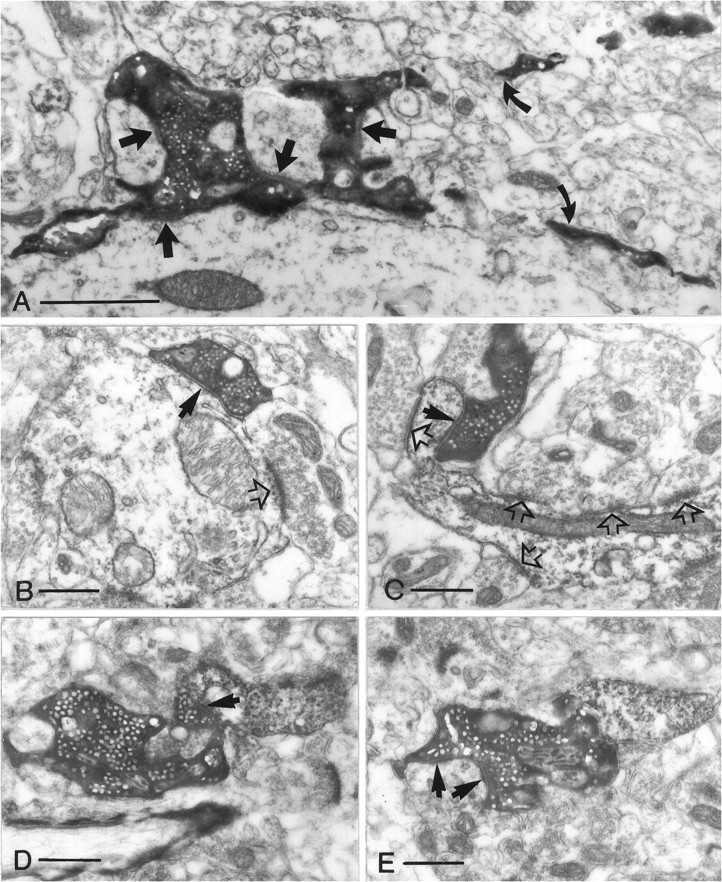
Terminal types of mossy fibers in the hilus. All electron micrographs have the same magnification. A, Mossy terminal establishing four synapses (arrows) with the shaft and the thorny excrescences of a mossy cell. Curved arrows point to the filopodiae originating from this terminal.B, A small en passant terminal with single release site (arrow) contacts a dendritic shaft.C, A drumstick-like small terminal forms a synapse on the proximal part of an SPR-positive spine. Open arrowsin B and C label synapses formed by unlabeled small terminals. D, E, Intermediate terminal type contacting a distal SPR-positive dendritic shaft (arrow in D) and on a neighboring section forming two synapses (arrows inE) on a putative mossy cell. Scale bars:A, 1 μm; B–E, 0.5 μm.