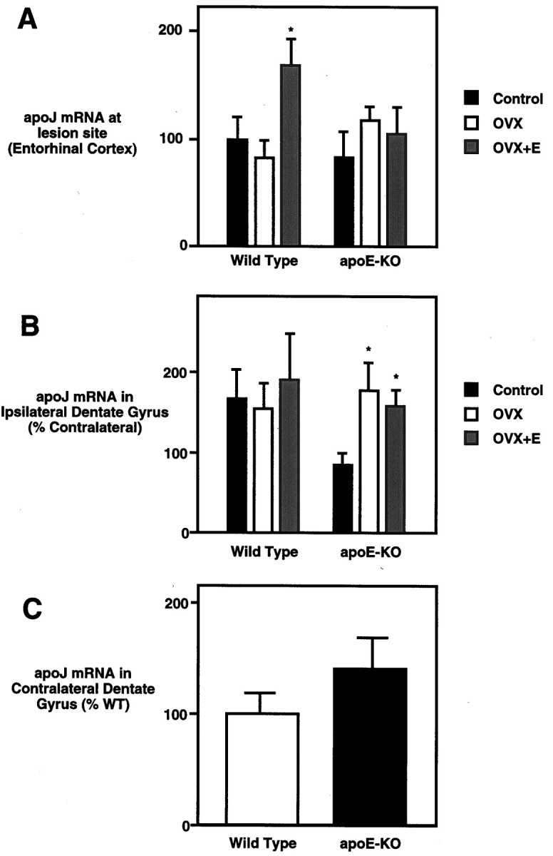
Fig. 3.

ApoJ mRNA levels show E2-dependent effects in wild-type but not ApoE-KO mice. A, At the lesion site (entorhinal cortex), apoJ mRNA level shows a nonsignificant decrease in response to OVX and a 1.7-fold increase with E2replacement. No significant changes were observed in apoE-KO mice. They-axis gives in situ grain density as a percent of WT, ovary-bearing (control) mice. B, In the deafferented dentate gyrus (ipsilateral to lesion), apoJ mRNA levels did not change in WT mice, whereas apoE-KO mice showed increased apoJ mRNA levels in response to ovariectomy. E2 replacement did not have an effect. Levels are given as a percent of contralateral values. The y-axis gives in situ grain density expressed as a percent of that in the contralateral (unlesioned) dentate gyrus of the same brain. C, In the unlesioned hippocampus (contralateral to lesion) of sham-ovariectomized mice, apoE-KO mice showed a nonsignificant increase in apoJ mRNA, suggesting a possible compensatory increase. This trend did not extend to the apoJ responses to E2 or lesion. They-axis gives the in situ grain density in the unlesioned dentate gyrus, expressed as a percent of that in WT mice. *Significantly different from sham-OVX mice;p < 0.05.
