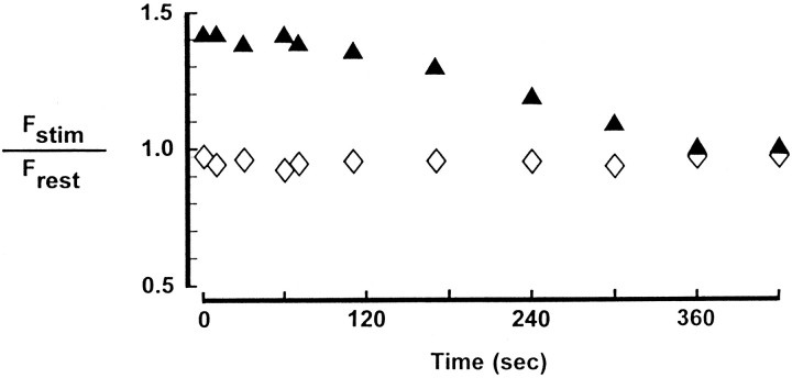
Fig. 1.
Ca Crimson fluorescence in larval neuromuscular preparations with and without nerve stimulation. Separate nerve terminals in a Ca Crimson-loaded preparation were analyzed for the relative intensity of fluorescence emission in the absence of nerve stimulation (⋄) or immediately after (at the times indicated) 2 min of 10 Hz stimulation of the motor nerve (▴). In the control preparation (⋄) the motor nerve of the adjacent hemisegment was stimulated (10 Hz, 2 min) to mimic the movement seen with stimulation of the correct motor nerve (▴). Frest was obtained from images captured 30 sec before the stimulus train.Fstim is mean bouton fluorescence assessed at the indicated times after stimulation. Data are mean values for at least 10 boutons per terminal. The SD did not exceed 20% of the mean.

