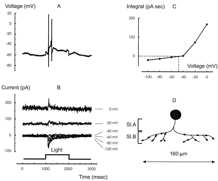
Fig. 3.
The light response, synaptic currents, and morphology of the narrow-field ON amacrine cells. A, The light response to a full-field red light stimulus. This cell type responds only at light ON. B, The current responses of the same cell to the same stimulus voltage clamped to the potentials indicated at the right of each trace. The inhibitory currents appear only at light ON. The light-evoked current reversed near −40 mV. C, The integral of the light-evoked current from 1000 to 2000 msec (ON response) plotted as a function of the holding potential. The curve, which was created by joining the points with linear segments, passes through zero at −48 mV, which indicates mixed excitatory and inhibitory inputs.D, Stylized sketch of the cell filled with 1% Lucifer yellow through a second electrode after electrical measurements. The cell ramified in sublamina B (Sl.B), consistent with its ON behavior. The extent of its processes was 160 μm, consistent with its classification as a narrow-field (100–200 μm) amacrine cell (Yang et al., 1991).

