
Fig. 4.
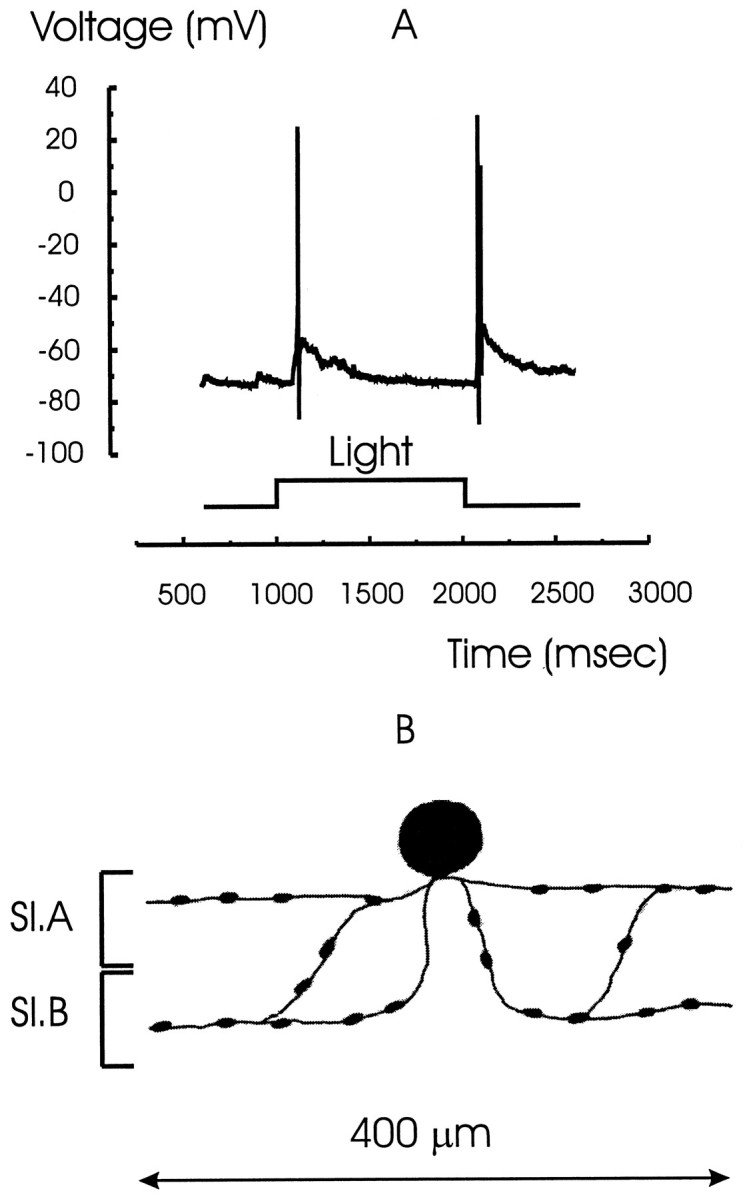
The light response and morphology of a wide-field amacrine cell. A, The light response of a wide-field amacrine cell that characteristically fires only one spike at light ON and OFF followed by a slow decay in response. B, Stylized sketch of the cell filled with Lucifer yellow. Processes ramify in both sublaminae A and B (Sl.A andSl.B) and spread laterally over ∼400 μm.