
Fig. 6.
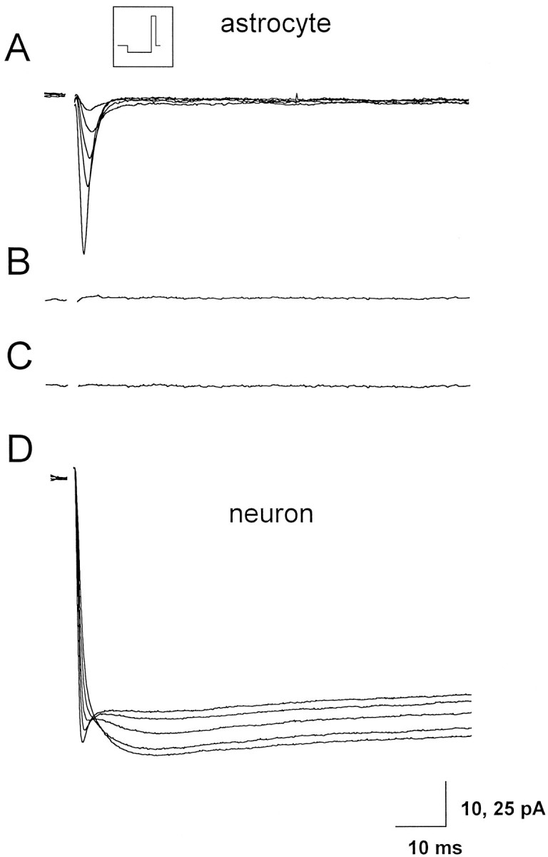
Whole-cell recordings of Ba2+and Na+ currents in hippocampal astrocytes and neurons. A, Progressive reduction of the Na+ current recorded from one astrocyte from the CA1 hippocampal region of a 10-d-old rat on slice perfusion with 10 mm BaCl2 and TEA in substitution of Na+ ions. The inset shows the pulse protocol used; the membrane was hyperpolarized to −110 mV from a holding potential of −80 mV for 0.5 sec and then depolarized to 0 mV for 100 msec. Successive voltage pulses were separated by a 4 sec interval. The capacitive transients have been blanked. LY included in the patch pipette diffused through gap junction from the recorded astrocyte into two other astrocytes (data not shown). B, No inward currents remain detectable after switching to Na+-free solution. C, Trace obtained after subtraction of the current traces recorded before and after 100 μm Cd2+ from the same astrocyte.D, Whole-cell recording from a CA1 pyramidal neuron of a 10-d-old rat with Ba2+ as charge carrier. Note the progressive increase of the Ba2+ current and the parallel reduction of the Na+ current on slice perfusion (which started immediately after establishing the whole-cell configuration) with 10 mm BaCl2 and TEA in substitution of Na+ ions. Stimulation protocol as inA.