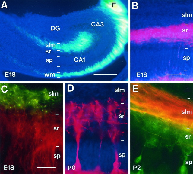
Fig. 1.
Layer-specific termination of developing connections in the hippocampus. A, Distribution of entorhinal fibers at E18 in the slm and in thewm, after an entorhinal injection of DiI.B, Distribution of associational fibers at E18 in thesr of the CA1 region after an injection of DiI in the CA3 region. C, E, Visualization of anterogradely traced entorhinal fibers and retrogradely labeled pyramidal cells in the CA1 region at E18 and P2, showing entorhinal afferents running above pyramidal cell dendrites. In C, entorhinal axons were labeled with DiA (green), and pyramidal neurons were labeled with DiQ (red); the reverse combination was used in E. D, Distribution of DiI-stained pyramidal cell apical dendrites at P0. Note that these dendrites terminate before the slm. Sections in A and B are counterstained with bisbenzimide. Scale bars: A, 250 μm; B, 100 μm; C–E, 50 μm. CA1,CA3, Hippocampal subfields; DG, dentate gyrus; F, fimbria; slm, stratum lacunosum-moleculare; sp, stratum pyramidale;sr, stratum radiatum; wm, white matter.

