
Fig. 11.
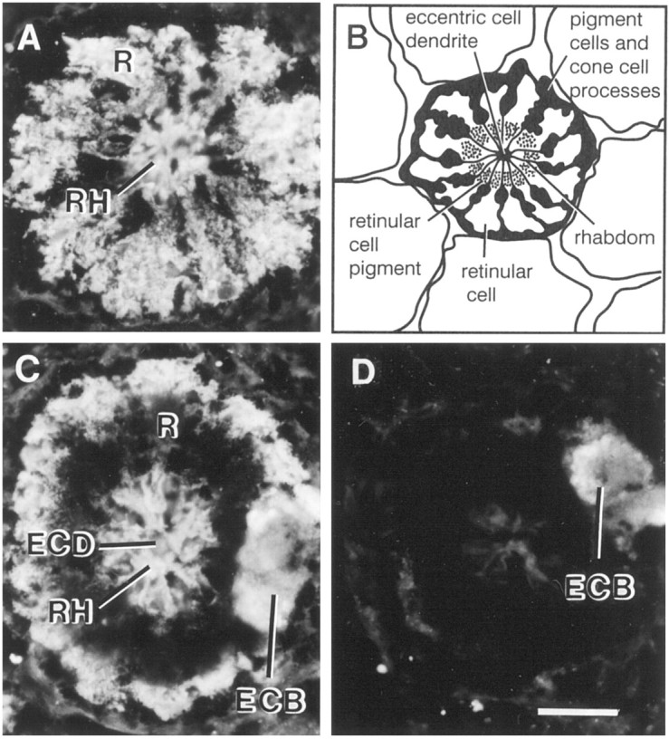
Localization of myoIIILimimmunoreactivity in fixed frozen sections of lateral eye. Lateral eyes dissected from light-adapted animals during the day were fixed in 4% paraformaldehyde as described in Materials and Methods. Frozen sections (14 μm) were incubated overnight in a 1:50 dilution of rat serum containing antibodies directed against the predicted C-terminal sequence of myoIIILim. The location of the primary antibody was visualized with a fluorescein isothiocyanate-conjugated secondary antibody. Specific myoIIILim is observed throughout the cytoplasm of the retinular cell and over the rhabdom. The staining observed over the eccentric cell body and dendrite is nonspecific.A, Cross-section of an ommitidium in the lateral eye at the level of the nuclei of the retinular cells. B, Diagram of a cross section of an ommitidium. The eccentric cell body is not shown. C, Cross-section of an ommatidium in the lateral eye at the level of the eccentric cell body. The dark region immediately peripheral to the rhabdom is occupied by pigment granules that absorb the fluorescent signal. D, A cross-section at a level similar to that shown in C was exposed to primary antibody that had been preincubated overnight with 10−5m free C-terminal peptide antigen.ECB, Eccentric cell body; ECD, eccentric cell dendrite; R, retinular cell; Rh, rhabdom. Scale bar, 40 μm.