
Fig. 3.
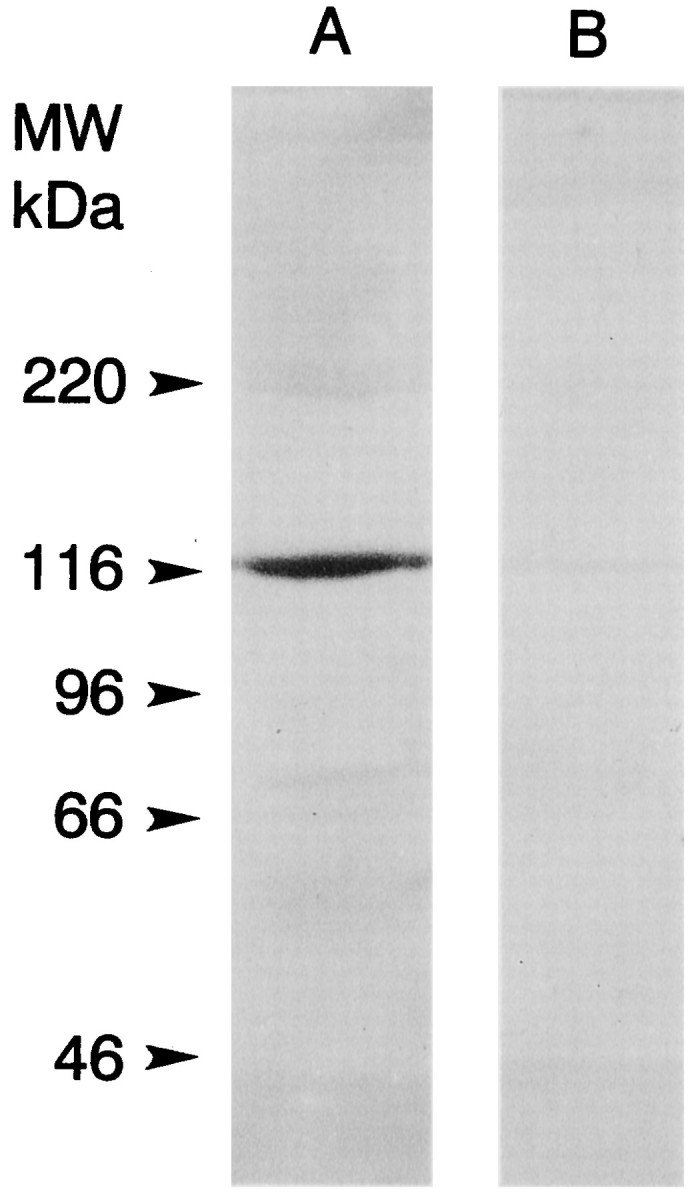
Western blots of soluble extracts of lateral eye and lateral optic nerve stained with an antibody directed against the predicted C terminus of myoIIILim. A soluble extract of lateral eye and lateral optic nerve (76 mg tissue wet weight) was prepared and concentrated as described in Materials and Methods for binding to calmodulin-Sepharose. The final volume of the concentrate was 200 μl. An aliquot was mixed 1:1 with 2× SDS sample buffer, and 5 μl/lane was fractionated by SDS-PAGE on a 7.5% gel and blotted to PVDF. Immunostaining was performed as described in Materials and Methods. The alkaline phosphatase-conjugated secondary antibody was used at a dilution of 1:2000. Lane A was incubated with a 1:100 dilution of serum from a rat injected with a peptide encoding the predicted C terminus of myoIIILim conjugated to keyhole limpet hemacyanin. A single immunostained band at ∼122 kDa was observed. Lane B was incubated with a 1:100 dilution of the same serum that had been preincubated overnight with 10−5m free C-terminal peptide. No immunostained bands were observed. The locations of the molecular mass standards are indicated.