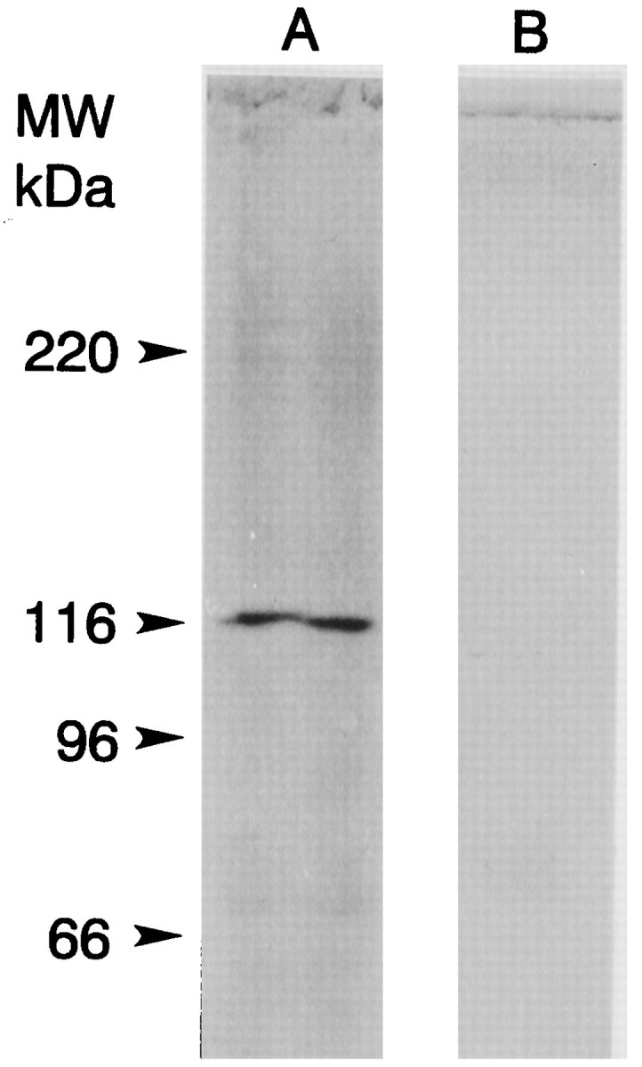
Fig. 7.

Western blot of a total protein extract of lateral eye showing myoIIILim-like immunoreactivity. Lateral eye tissue was homogenized in HB (20 μl/mg tissue wet weight) (Edwards and Battelle, 1987), and then the homogenate was diluted 1:1 with 2× SDS sample buffer and sonicated. Ten microliters of the SDS-solubilized protein were applied to the lanes. Immunostaining was performed as described in Materials and Methods and Figure 3. Lane Awas incubated with a 1:100 dilution of ascites fluid from a mouse that had been immunized with gel-purified 122 kDa myoIIILim.Lane B was incubated with a 1:100 dilution of ascites from an unimmunized mouse. The locations of the molecular mass markers are indicated. A single immunostained band at 122 kDa is seen in the lane incubated with antibody directed against the 122 kDa myoIIILim. No immunostained bands with higher molecular mass were detected.
