
Fig. 2.
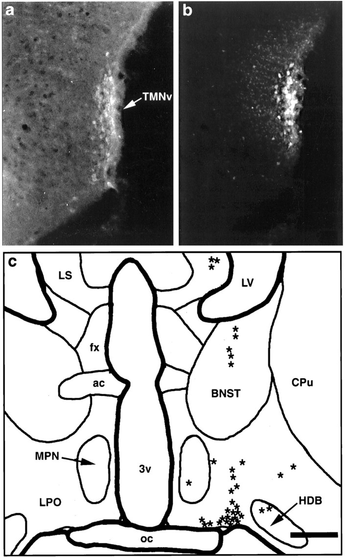
Summary of findings from a fluorogold injection into the core of the TMNv with no involvement of adjacent structures. a, Fluorescence photomicrograph of a caudal hypothalamic section stained immunocytochemically for adenosine deaminase, as visualized using a rhodamine filter cube. Immunoreactive neurons delineate the TMNv (arrow). b, Fluorescence photomicrograph of the same field, demonstrating Fluorogold fluorescence as seen through a UV filter cube, showing that the center of the injection is limited to and essentially demarcates the TMNv. c, Camera lucida drawing of a caudal preoptic section showing retrogradely labeled cells (eachasterisk represents one cell) produced by the injection in b. In this case, the vast majority of retrogradely labeled neurons were concentrated in the ventral portion of the lateral preoptic area. This pattern of labeling extended roughly 300 μm rostrally and 100 μm caudally from the level drawn. Scale bar (shown in c): a, b, 400 μm; c, 800 μm.