
Fig. 4.
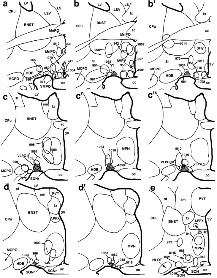
A summary diagram illustrating biotinylated dextran injection sites at six levels of the preoptic area. The VLPO is most prominent in schematics c, c′,d, and d′ (in light gray).Asterisks denote cases VLPO 11, VLPO 38, and R 1059 in which biotinylated dextran injections were predominantly located within the VLPO (see Results).